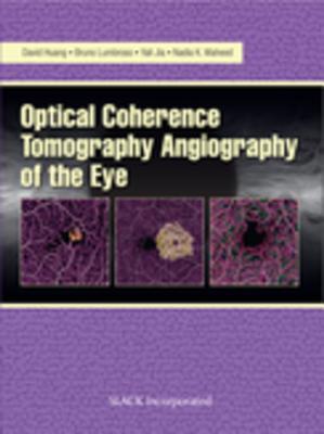
Full Download Optical Coherencre Tomography Angiography of the Eye - David Huang | PDF
Related searches:
Optical coherence tomography - OCT - The Eye Practice
Optical Coherencre Tomography Angiography of the Eye
Optical Coherence Tomography - The Cardiology Advisor
Optical coherence tomography (oct) is a procedure used for the noninvasive examination of intraocular structures. Primarily used for analysis of the retina and optic nerve, oct centers on the amount of light absorption or scattering that occurs when light passes through a given tissue layer.
Optical coherence tomography has become established as an imaging modality in clinical ophthalmology and is finding applications in even gastrointestinal and dermatological arenas. Its biggest application in cardiovascular medicine currently is in the coronary vasculature and to a lesser extent coronary graft assessment.
Optical coherence tomography (oct) is a non-invasive diagnostic technique that renders an in vivo cross sectional view of the retina. Oct utilizes a concept known as inferometry to create a cross-sectional map of the retina that is accurate to within at least 10-15 microns.
Optical coherence tomography (oct) is useful in diagnosing many eye conditions. Oct is often used to evaluate disorders of the optic nerve as well. Optical coherence tomography (oct) is useful in diagnosing many eye conditions. Oct is often used to evaluate disorders of the optic nerve as well.
Optical coherence tomography (oct) is an imaging technique that provides high resolution, non-destructive, in situ, real-time reflectivity profiling of non-absorptive samples. Medical applications include noninvasive ophthalmological imaging and endoscopic gastrointestinal tract imaging.
Introduction optical coherence tomography, or oct is a non- contact, noninvasive imaging technique used to obtain high resolution 10 cross sectional images of the retina and anterior segment.
Optical coherence tomography (oct) is a micron-scale imaging method, analogous to ultrasound measuring the backreflection of infrared light rather than sound. It can operate in 2d and 3d mode and exceeds the frame rate of video. The penetration depth of oct is about 2 mm, depending on tissue types.
Optical coherence tomography (oct) is the optical analog of ultrasound imaging and is a powerful imaging technique that enables non-invasive, in vivo, high resolution, cross-sectional imaging in biological tissue. Between 30 to 40 million oct imaging procedures are performed per year in ophthalmology.
European space agency astronaut alexander gerst, expedition 40 flight engineer, uses the optical coherence tomography (oct) camera during an ocular health (oh) vision test in the harmony node of the international space station. The oh experiment observes and seeks to understand vision changes during long-term space missions.
Optical coherence tomography of ocular diseases, third edition is written with the clinician in mind. The text’s primary objective is to illustrate the appearance of the eye in health and disease, comparing conventional clinical technologies using spectral domain oct imaging.
Spectroscopic optical coherence tomography (soct) is an optical imaging and sensing technique, which provides localized spectroscopic information of a sample based on the principles of optical coherence tomography (oct) and low coherence interferometry.
Optical coherence tomography 3d oct-1 (type:maestro2) smart infrastructure.
Optical coherence tomography – or oct – is a medical imaging technique. It uses visible light to produce a 3-d image of what’s happening beneath the surface.
Optical coherence tomography (oct) is a diagnostic procedure that is used during cardiac catheterization. Unlike ultrasound, which uses sound waves to produce an image of the blood vessels, oct uses light. With oct, doctors can obtain images of the blood vessels that are about the same as if they were looking under a microscope.
Optical coherence tomography (oct) is a noninvasive imaging technology used to obtain high-resolution cross-sectional images of the retina. Oct is similar to ultrasound testing, except that imaging is performed by measuring light rather than sound.
A technique called optical coherence tomography (oct) has been developed for noninvasive cross-sectional imaging in biological systems. Oct uses low-coherence interferometry to produce a two-dimensional image of optical scattering from internal tissue microstructures in a way that is analogous to ultrasonic pulse-echo imaging.
Optical coherence tomography optical coherence tomography (oct) is a non-invasive way for your physician to visualize the microscopic structures of your retina. The technology has revolutionized our care for patients with conditions affecting the retina, such as macular degeneration, diabetic retinopathy, retinal vein occlusions, and many more.
This guide was created to provide information to students, residents and clinicians in eye care to better understand optical coherence tomography scans. Examples are provided to help understand optic nerve and retinal pathologies to assist in the diagnosis and management of these conditions.
Optical coherence tomography (oct) is a sensitive, noninvasive test used to create high-resolution images of the central retina (macula). Oct uses light (not x-ray) that is shone onto and reflected by the different layers of the retina to measure thickness and to detect the extent of various conditions which affect the macula.
Optical coherence tomography a test used to follow patients diagnosed with ms on their recovery and response to treatments.

Post Your Comments: